PRISMA by Omica
A unified digital workspace for clinical pathology.
PRISMA brings whole-slide imaging, clinical context, synoptic reporting, and on-slide AI analysis into a single sign-out environment. Deployed on-premise in your laboratory.


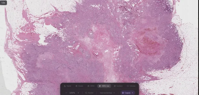
PRISMA workspace. The HER2-low inference layer reflects 41,696 evaluated patches. Case details are illustrative.
The workspace
All diagnostic context beside the slide.
Eliminate context switching between separate LIS terminals, image viewers, clinical guidelines, and report templates. PRISMA unifies case management, high-resolution viewing, and diagnostic tools in one interface.
-
Fig 0.1
Case management
Cases ingest directly from the LIS with associated whole-slide images. Prioritize worklists, review, and sign out in a single workflow that syncs back to the LIS.
-
Fig 0.2
Embedded AI analysis
Nuclear segmentation, tumor boundaries, histologic grade, and biomarker predictions run at scan time and display as verified diagnostic layers.
-
Fig 0.3
Morphologic retrieval
Search historical archives by histologic pattern to reference verified index cases beside your active read.
-
Viewer
Whole-slide images at full optical resolution, multi-slide case navigation, and persistent diagnostic annotations.
-
Guidelines in context
Standardized clinical guidelines open adjacent to the slide, keeping diagnostic criteria in view during review.
-
Agent
Query historical cases, retrieve relevant staging criteria, or cross-reference previous pathology reports in plain language.
-
Synoptic reporting
Structured synoptic templates populated directly from slide review and verified AI outputs.
Diagnostic AI models
A library of diagnostic AI models, running on-premise.
PRISMA executes validated AI models locally across tissue types and stains. Review nuclear density, tumor margins, growth patterns, grade, and predictive biomarker status as interactive overlays.
| Organ | Cell counts | Tumor detection | Tissue map | Pattern and grade | Immune infiltrate | Biomarkers |
|---|---|---|---|---|---|---|
| Breast | ||||||
| Lung | Not available | Not available | Not available | |||
| Prostate | Not available | Not available | Not available | |||
| Colorectal | Not available | Not available | ||||
| Lymph node | Not available | Not available | Not available | Not available | ||
| Pancreas | Not available | Not available | Not available |
Breast · Biomarkers
HER2, HER2-low, and molecular subtype predicted from the slide.
Brightfield H&E
Comprehensive nuclear segmentation, tissue segmentation, grading, and biomarker prediction on standard H&E.
Brightfield IHC (H-DAB)
Nuclear detection, membrane quantification, and positive cell scoring.
Fluorescence and multiplex
Cellular and subcellular segmentation across multiplexed immunofluorescence panels.
PRISMA diagnostic models provide assistive analytical layers for review by pathologists. Analytical outputs do not constitute a standalone diagnosis. Model availability varies by tissue indication, stain, and jurisdiction.
Deployment options
Deployment models for any laboratory stage.
Select the deployment path that fits your existing infrastructure, or combine them to transition historical and prospective workflows.
-
Path 01
Connect existing scanners
PRISMA supports leading digital pathology scanners including Hamamatsu, Aperio, Leica, and Omica Edge-40 systems. Ingest DICOM WSI, SVS, NDPI, and BigTIFF directly into your LIS workflow.
-
Path 02
Deploy Nucleo on-premise
Nucleo is Omica's on-premise compute and scanning appliance. It executes PRISMA and GPU-accelerated inference locally, ensuring patient health information and slide data remain within your hospital firewall.
Request a Nucleo walkthrough -
Path 03
Turnkey archive digitization
Omica manages high-throughput scanning of historical slide archives, either through secure logistics or dedicated on-site scanning units, indexing all glass directly into PRISMA.
Inquire about archive digitization
Recommended archive scope: Prioritize recent surgical resections and high-risk biopsies most relevant for recurrence monitoring and clinical review.
Clinical network
Connected clinical pathology networks.
Coordinate inter-institutional secondary consults digitally, contribute consented cases to oncology research through the Omica Cancer Atlas, and participate in multi-center biomarker screening programs.
Implementation
Frequently asked questions
What is PRISMA?
PRISMA is Omica's digital pathology workspace. It unifies whole-slide viewing, case accession, synoptic reporting, and local AI model execution into a single environment. PRISMA deploys on-premise via Nucleo or certified server infrastructure within your institution.
Do we need to purchase new scanning hardware?
No. PRISMA integrates directly with your existing scanners, or you can deploy Nucleo for integrated scanning and compute. Alternatively, Omica provides turnkey archive digitization on-site or through secure logistics.
Which scanners and file formats are supported?
PRISMA supports leading digital pathology scanners including Omica Edge-40, Hamamatsu, Aperio, and Leica. Supported formats include DICOM WSI, SVS, NDPI, and BigTIFF.
Does any patient data leave our network?
No. Slide images, diagnostic reports, and AI inference outputs remain entirely within your institutional network firewall on local storage and compute.
How does PRISMA integrate with hospital LIS and PACS?
PRISMA provides bi-directional integration with laboratory information systems via HL7 and FHIR, and interfaces with institutional PACS and EHR systems for frictionless order and result exchange.
Does the AI make the diagnosis?
No. PRISMA's AI tools provide assistive analytical layers for review by board-certified pathologists. The pathologist retains complete diagnostic discretion and signs out each case. Regulatory clearances vary by model, tissue indication, and jurisdiction.